
How Does The Skeletal System Work With The Muscular System
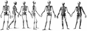
The Skeletal System
The skeletal system comprises 206 bones. Of the 206, six of them are ossicles located in the ear. Infants have about 350 bones. However, most will eventually connect, forming the 206 bones in adults. The bones provide a solid framework, supporting and shaping the body. They protect delicate or soft parts and anchor muscles.
 Bones are osseous cells hardened by calcium and phosphorus from our food. The periosteum is a membrane that covers every bone. It is filled with blood vessels carrying bone-building materials and minerals to the cells to keep them alive and hardened by filling in the spaces between them.
The bones of a baby are partly cartilage, which is bone cells not fully hardened. Bones are not wholly hardened until a person is about twenty years old. This is why adults can break their bones easier than children. Bone cells multiply rapidly during the growing years but slowly after that as bones need to be repaired. With age, bones become more rigid and brittle.
Long bones—The shaft, or diaphysis, is the lengthy section that is rigid and compact. The end, or epiphysis, is sponge-like and covered by a shell of more rigid bone. The shaft and end do not fuse until total growth is achieved.
Joints—These are where the bones are attached. They enable you to move your legs and arms. Fibrous, strong bands, called ligaments, hold them together. The moving joints are lined with a membrane that secretes synovial fluid. This keeps the joints “oiled” and working smoothly. Cartilage plates on the ends of bones make a slick surface for rotation.
Types of joints—Finger joints move like a door on hinges and are called hinge joints. The hips and shoulders are ball-and-socket joints that allow a rotating movement. The rounded end of one bone fits into the hollowed-out end of the other.
Joint problems—Arthritis and rheumatoid arthritis occur in the joints. Other types include tubercular (TB) arthritis, gonorrheal arthritis, and gout. Synovitis is swelling of the lining of the joint. (Rheumatism is an inflammation in the muscles.
Vertebral column—Your spine supports your head, stiffens the mid-portion of your body, and anchors your ribs and pelvic bones. It also shields the spinal cord, which passes down through its bony rings from the brain. The spine also carries the body weight and consists of twenty-six irregularly shaped bony rings called vertebrae (vertebra is singular). On the internal side of each vertebra is a bony structure called the arch, which forms an opening or spinal foramen through which the spinal cord passes.
Several finger-like extensions or processes on which ligaments and tendons are anchored protrude from the arch. The discs are cartilage plates between these rings, which are shock absorbers when you walk, sit, or fall. They make the joints flexible so that you can turn and bend.
Spinal curves—The spine has four standard curves. Disease, injury, or poor posture distort these curves, producing kyphosis (hunchback), lordosis (swayback), or scoliosis (lateral curvature).
The seven top vertebrae (in your neck) are the cervicals. The next twelve in your upper back are the thoracic. The following five below are the lumbers in your lower back. Then come five sacral vertebrae and four coccygeal in your so-called “tailbone.” Without that section, you could not sit properly.
Ribs—Your rib cage is formed by twelve ribs on each side of a central divide. The sternum is the breast bone, a flat bone in the upper center of your chest. The upper seven pairs of ribs are connected to the sternum in front. The following three pairs are connected to the sternum in front. The successive three pairs are attached indirectly to the sternum. The last two are free in front and are called floating ribs. They help your chest expand as you inhale.
Pelvic bones—Your pelvic girdle is formed from several bones. In women, the central opening is called the birth canal. Babies could not be born if they were not more significant than men.
Leg bones—The femur is the upper bone of the leg and is the longest and strongest bone in your body; its upper end is attached to the pelvic bone in a ball-and-socket joint. Try to avoid falls, which could break that joint, called “breaking the hip.” The lower legs have two bones: the tibia, or shin bone, in front and the fibula in the back. The knee cap (patella) covers the knee joint so you can kneel.
Ankle and foot bones—There are seven tarsal bones in the ankle, with the largest in the heel. They join the five metatarsals (instep bones) to form two arches—one lengthwise across the foot (the longitudinal arch) and the other from side to side (the metatarsal arch). Weak muscles reduce the “spring,”; and flat feet can result. The fourteen bones of the toes, the phalanges, are attached to the metatarsal bones. The large toe has two phalanges; each of the others is three.
Shoulder bones—In front are two long, thin bones—the clavicles or collarbones. In the back, there is the two scapula (scapulars) or shoulder blades. Because all these bones are only attached to the sternum, you can freely move your shoulders and arms up, down, forward, and back.
Arm bones—The humerus is the single long bone in the upper arm. The upper end is connected to the scapula: and the lower end connects with the ulna to form the elbow joint (what people call your “funny bone”). There are two bones in the forearm. The more significant is the ulna and the other radius. Because God gave you two bones in your forearm, you can rotate your hand in a semicircle instead of one. As you extend your hand with the thumb up, the radius is on top and is the one that rotates.
Hand bones—There are eight carpal or wrist bones. Next come five metacarpal bones, forming the palm of your hand; then come to the phalanges (finger bones). There are three phalanges on each finger and two on the thumb. You have twenty-nine joints in each wrist and hand to produce fine movements.
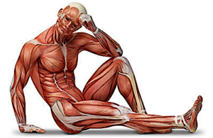
Muscular System Function
Without muscles, you could not move an inch. Muscles are connected to bones by tendons, which enable them to move the bones. Muscle cells are tiny elastic threads of protein wrapped in bundles; several bundles make a muscle. Each muscle is covered by a covering of connective tissue called fascia, the ends of which lengthen into tough cords called tendons, which are attached to bones.
 Bursas—The tendons have sheaths lined with synovial membranes that allow smooth, gliding movement. Bursas (bursae) are tiny sacs with synovial membranes found wherever pressure is exerted over moving parts. When bursae become inflamed, bursitis is the result. This occurs in the knee joint and is called “housemaid’s knee.” Muscles work in pairs. Nerves in the muscle bundles direct movements, and blood vessels carry food materials—an overstretched or damaged muscle results in pain.
Flexion and extension—When you bend your arm. Your arm muscle becomes shorter and thicker; that is flexion. When you straighten it out, the muscles lengthen and narrow; that is extension. A muscle is most potent when contracted. Keeping your arm muscles close to your body and your back straight when you move something makes you less likely to overstrain. Pick things up with your muscles, not your spine.
Power—Muscles use oxygen and a type of sugar to provide the power to do things. The result is energy and heat. In fact, muscles produce most of the body heat (with the liver in second place as a heat source). Using muscles builds them up. Otherwise, they become flabby.
Waste products—Two waste products are produced when a muscle works. Carbon dioxide is carried to the lungs, while lactic acid is removed through the kidneys and sweat glands. If you work too hard, so much lactic acid remains in the muscles that they ache and feel sore afterward.
Rehabilitation—An injured or inactive muscle can be retrained to do its work. This must be done slowly and carefully. The person you are helping must be encouraged to persevere.
Control—There are two types of muscles you consciously control the voluntary (skeletal) muscles. The involuntary (smooth) muscles work automatically. These latter muscles include the kidneys, heart, stomach, etc.
Breathing muscles—Of the 325 muscles in the body, some are essential for breathing. The diaphragm is between the abdominal and chest cavities and helps you breathe. Muscles between the ribs, and the intercostals, also help enlarge the chest cavity.
Abdominal muscles—These are mentioned because they are the most likely to produce hernias. They are arranged to give support by overlapping layers from various angles. There are also unique places where rupture (hernia) with protrusion of part of the intestine may occur. These weak places are where blood vessels, nerves, ligaments, and cords pass through the muscles. The inguinal rings, the femoral rings, and the umbilicus are common sites for hernias.
Shoulders and buttocks—These muscles are essential if you need to give intramuscular injections to someone. In addition, the shoulder muscles sometimes become chilled, and aches develop.
Leg muscles—The Achilles tendon is the large tendon that attaches the calf muscle to the heel bone. Sometimes it becomes strained or injured.